Research progress in the influencing factors and correction methods of XRF-CS
HUANG Ping-An1,2(), WANG Xia-Qing2(), TANG Xiang-Ling1, WANG Yu-Tang1,2, LI Wei2, LUO Zeng2, Lyu Fei-Ya2
1. College of Earth Science, Guilin University of Technology, Guilin 541004, China 2. College of Geography and Tourism, Hunan University of Arts and Science, Changde 415000, China
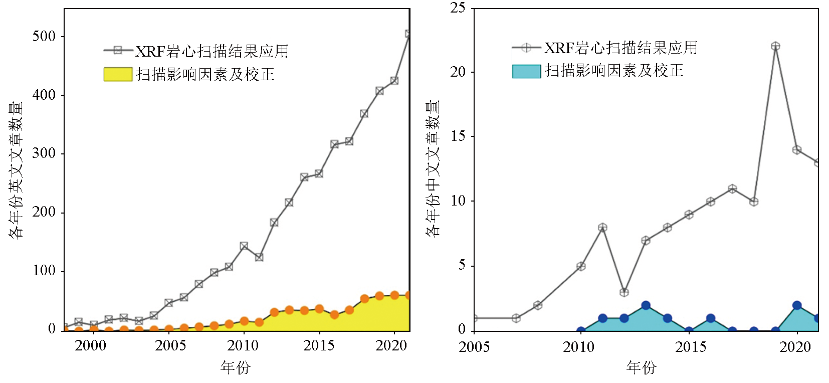
With more than 20 years of development, the X-ray fluorescence core scanners (XRF-CS) have been widely applied in the elemental analysis of multi-type sediment cores, the paleoenvironment reconstruction, and the exploration of mineral reservoirs and their abundance, exhibiting great potential for application. However, there is a lack of studies on the influencing factors and correction of the elemental signals output by XRF-CS (especially in China), which restricts the proper use of XRF-CS and the accurate interpretation of their data. Compared with conventional XRF techniques, XRF-CS enjoy a high processing speed (only 1/10 of the time for conventional analysis), high continuity, non-destructive scanning, and a high resolution (up to 0.02 mm). However, XRF-CS only output semi-quantitative values of elemental signals and thus fail to accurately identify the element compositions. This study summarized the influencing factors of the values of the elemental signals output by XRF-CS in terms of instruments and cores, together with the degrees of the influences. On this basis, this study proposed achieving the balance between the intensity of elemental signals output by XRF-CS and cost by selecting appropriate scanning steps and exposure time on the premise of the optimal instrument setting. This study also suggested that the influences of water content and particle sizes on elemental signals should be eliminated as far as possible by drying in the air and smoothing the core surface during the scanning. To improve the accuracy of elemental signals output by XRF-CS, this study systematically introduced three types of international common calibration models and their application potential, namely the normalized median-scaled (NMS) model, the log-ratio calibration equation (LRCE) model, the improved multivariate log-ratio calibration (MLC) model, the normalized polynomial-scaled calibration (NPS) model, and polynomial-corrected multivariate log-ratio calibration (P-MLC) model. Finally, this study proposed further enhancing research on the comparative analysis of the influence exerted by the same factor among multiple types of XRF-CS; the optimization of calibration models and development of visual software packages; the equipment of multiple sensors for integrated scanning, and the extensive applications in the exploration and evaluation of geological and mineral resources.
黄平安, 王夏青, 唐湘玲, 王玉堂, 李玮, 罗增, 吕飞亚. X射线荧光光谱岩心扫描影响因素及校正方法的研究进展[J]. 物探与化探, 2023, 47(3): 726-738.
HUANG Ping-An, WANG Xia-Qing, TANG Xiang-Ling, WANG Yu-Tang, LI Wei, LUO Zeng, Lyu Fei-Ya. Research progress in the influencing factors and correction methods of XRF-CS. Geophysical and Geochemical Exploration, 2023, 47(3): 726-738.
Yu J M, Oppo D W, Jin Z D, et al. Millennial and centennial CO2 release from the Southern Ocean during the last deglaciation[J]. Nature Geoscience, 2022, 15:293-299.
doi: 10.1038/s41561-022-00910-9
[2]
Wilhelm B, Rapuc W, Amann B, et al. Impact of warmer climate periods on flood hazard in the European Alps[J]. Nature Geoscience, 2022, 15(2):118-123.
doi: 10.1038/s41561-021-00878-y
Liao S L, Tao C H, Zhao J N, et al. Application of PXRF in sediment analysis for geochemical prospecting in Dragon Horn area on the southwestern Indian Ridge[J]. Bulletin of Geological Science and Technology, 2022, 41(3):264-272.
[4]
Jansen J H F, Van der Gaast S J, Koster B, et al. CORTEX,a shipboard XRF-scanner for element analyses in split sediment cores[J]. Marine Geology, 1998, 151(1-4):143-153.
doi: 10.1016/S0025-3227(98)00074-7
[5]
Yi L, Wang H F, Deng X G, et al. Geochronology and geochemical properties of Mid-Pleistocene sediments on the Caiwei Guyot in the Northwest Pacific imply a surface-to-deep linkage[J]. Journal of Marine Science and Engineering, 2021, 9(3):253.
doi: 10.3390/jmse9030253
[6]
Gregory B R B, Patterson R T, Galloway J M, et al. The impact of cyclical,multi-decadal to centennial climate variability on arsenic sequestration in lacustrine sediments[J]. Palaeogeography,Palaeoclimatology,Palaeoecology, 2021, 565:110189.
doi: 10.1016/j.palaeo.2020.110189
[7]
Guo F, Clemens S C, Wang T, et al. Monsoon variations inferred from high-resolution geochemical records of the Linxia loess/paleosol sequence,western Chinese Loess Plateau[J]. Catena, 2021, 198:105019.
doi: 10.1016/j.catena.2020.105019
[8]
Henares S, Bloemsma M R, Donselaar M E, et al. The role of detrital anhydrite in diagenesis of aeolian sandstones (upper Rotliegend,the Netherlands):Implications for reservoir-quality prediction[J]. Sedimentary Geology, 2014, 314:60-74.
doi: 10.1016/j.sedgeo.2014.10.001
Wu L J, Li G. The estimation of organic contents in marine sediments based on bromine intensity by the XRF scanner[J]. Journal of Tropical Oceanography, 2022, 41(2):112-120.
doi: 10.11978/2021041
Zhang Y Z, Zhang J W, Mao C H, et al. Accuracy assessment and calibration of the impact of water content and structure of lake sediments on the XRF scanning data—A case study of Aweng Co in the Tibetan Plateau[J]. Quaternary Sciences, 2020, 40(5):1145-1153.
[11]
Tjallingii R, Röhl U, Kolling M, et al. Influence of the water content on X-ray fluorescence core-scanning measurements in soft marine sediments[J]. Geochemistry,Geophysics,Geosystems, 2007, 8(2):Q02004.
[12]
Richter T O, Van der Gaast S, Koster B, et al. The Avaatech XRF core scanner:Technical description and applications to NE Atlantic sediments[J]. Geological Society of London Special Publication, 2006, 267:39-50.
doi: 10.1144/GSL.SP.2006.267.01.03
[13]
Croudace I W, Rindby A, Rothwell R G. ITRAX:Description and evaluation of a new multi-function X-ray core scanner[J]. Geological Society of London Special Publication, 2006, 267:51-63.
doi: 10.1144/GSL.SP.2006.267.01.04
[14]
Hennekam R, de Lange G. X-ray fluorescence core scanning of wet marine sediments:Methods to improve quality and reproducibility of high-resolution paleoenvironmental records[J]. Limnology and Oceanography:Methods, 2012, 10:991-1003.
doi: 10.4319/lom.2012.10.991
[15]
Zuo R G. ITRAX:A potential tool to explore the physical and chemical properties of mineralized rocks in mineral resource exploration[J]. Journal of Geochemical Exploration, 2013, 132:149-155.
doi: 10.1016/j.gexplo.2013.06.010
[16]
Halim A Y, Kelloway S J, Marjo C, et al. A Hylogger-Itrax core-scanner comparison for multi-scale high-resolution petrophysical characterisation workflow[J]. Applied Geochemistry, 2021, 133:104956.
doi: 10.1016/j.apgeochem.2021.104956
[17]
Haschke M. The Eagle III BKA system,a novel sediment core X-ray fluorescence analyser with very high spatial resolution[J]. Geological Society of London Special Publication, 2006, 267:31-37.
doi: 10.1144/GSL.SP.2006.267.01.02
Boyle J F, Chiverrell R C, Schillereff D. Approaches to water content correction and calibration for μXRF core scanning:Comparing X-ray scattering with simple regression of elemental concentrations[G]//Croudace I W,Rothwell R G.Micro-XRF studies of sediment cores:Applications of a non-destructive tool for the environmental sciences.Dordrecht,Netherlands:Springer, 2015:373-390.
Chen Y L, Zheng H B. The application of XRF core scanning to Quaternary sediments[J]. Marine Geology Frontiers, 2014, 30(4):51-59.
[21]
Mondal M N, Horikawa K, Seki O, et al. Investigation of adequate calibration methods for X-ray fluorescence core scanning element count data:A case study of a marine sediment piston core from the Gulf of Alaska[J]. Journal of Marine Science and Engineering, 2021, 9(5):540.
doi: 10.3390/jmse9050540
[22]
Nowaczyk N R, Liu J B, Plessen B, et al. A high-resolution paleosecular variation record for marine isotope stage 6 from Southeastern Black Sea sediments[J]. Journal of Geophysical Research:Solid Earth, 2021, 126(3): e2020JB021350.
[23]
Hansen K E, Giraudeau J, Limoges A, et al. Characterization of organic matter in marine sediments to estimate age offset of bulk radiocarbon dating[J]. Quaternary Geochronology, 2022, 67:101242.
doi: 10.1016/j.quageo.2021.101242
[24]
Johnson J E, Phillips S C, Clyde W C, et al. Isolating detrital and diagenetic signals in magnetic susceptibility records from methane-bearing marine sediments[J]. Geochemistry,Geophysics,Geosystems, 2021, 22(9): e2021GC009867.
[25]
Lyle M, Lyle A O, Gorgas T, et al. Data report:Raw and normalized elemental data along the Site U1338 splice from X-ray fluorescence scanning[J]. Proceedings of the Integrated Ocean Drilling Program, 2012, 320/321:1-19.
Zhang X L, Fan D J, Wang L, et al. The calibration and quality evaluation of elemental analysis results of marine sediment measured by an X-ray fluorescence core scanner[J]. Acta Oceanologica Sinica, 2013, 35(6):86-95.
[27]
Shackford J K, Lyle M, Wilkens R H, et al. Data report:Raw and normalized elemental data along the Site U1335,U1336,and U1337 splices from X-ray fluorescence scanning[J]. Proceedings of the Integrated Ocean Drilling Program, 2014, 320/321:1-19.
[28]
Lefebvre P, Sabatier P, Mangeret A, et al. Climate-driven fluxes of organic-bound uranium to an alpine lake over the Holocene[J]. Science of the Total Environment, 2021, 783:146878.
doi: 10.1016/j.scitotenv.2021.146878
[29]
Hagemans K, Nooren K, de Hass T, et al. Patterns of alluvial deposition in Andean lake consistent with ENSO trigger[J]. Quaternary Science Reviews, 2021, 259:106900.
doi: 10.1016/j.quascirev.2021.106900
[30]
Zhao Y T, An C B, Zhou A F, et al. Late Pleistocene hydroclimatic variabilities in arid north-west China:Geochemical evidence from Balikun Lake,eastern Tienshan,China[J]. Journal of Quaternary Science, 2021, 36(3):415-425.
doi: 10.1002/jqs.v36.3
[31]
Knierzinger W, Huang J J S, Strasser M, et al. Late Holocene periods of copper mining in the Eisenerz Alps (Austria) deduced from calcareous lake deposits[J]. Anthropocene, 2021, 33:100273.
doi: 10.1016/j.ancene.2020.100273
Yang H F, Zhao Y, Cui Q Y, et al. Paleoclimatic indication of X-ray fluorescence core-scanned Rb/Sr ratios:A case study in the Zoige Basin in the eastern Tibetan Plateau[J]. Science China Earth Sciences, 2021, 51(1):73-91.
Cui Q Y, Zhao Y. Climatic abrupt events implied by lacustrine sediments of Arxan Crater Lake,in the central Great Khingan Mountains,NE China during Holocene[J]. Quaternary Sciences, 2019, 39(6):1346-1356.
Fan P F, Deng S P, Zou Y, et al. Application of XRF semi-quantitative analysis technology in identifying ore on polished section[J]. Journal of Jinlin University:Earth Science Edition, 2021, 51(3):783-791.
[35]
Wang X Q, Wang Z S, Xiao J, et al. Soil erosion fluxes on the central Chinese Loess Plateau during CE 1811 to 1996 and the roles of monsoon storms and human activities[J]. Catena, 2021, 200:105148.
doi: 10.1016/j.catena.2021.105148
[36]
Sun Y B, Clemens S C, Guo F, et al. High-sedimentation-rate loess records:A new window into understanding orbital- and millennial-scale monsoon variability[J]. Earth-Science Reviews, 2021, 220:103731.
doi: 10.1016/j.earscirev.2021.103731
[37]
Wang X Q, Jin Z D, He Z, et al. New insights into dating the sediment sequence within a landslide-dammed reservoir on the Chinese Loess Plateau[J]. The Holocene, 2019, 29(6):1020-1029.
doi: 10.1177/0959683619831426
[38]
Sun Y B, Liang L J, Bloemendal J, et al. High-resolution scanning XRF investigation of Chinese loess and its implications for millennial-scale monsoon variability[J]. Journal of Quaternary Science, 2016, 31(3):191-202.
doi: 10.1002/jqs.v31.3
[39]
Wang X Q, Jin Z D, Zhang X B, et al. High-resolution geochemical records of deposition couplets in a palaeolandslide-dammed reservoir on the Chinese Loess Plateau and its implication for rainstorm erosion[J]. Journal of Soils and Sediments, 2018, 18(3):1147-1158.
doi: 10.1007/s11368-017-1888-9
[40]
Wang X Q, Jin Z D, Chen L M, et al. High-resolution X-ray fluorescence core scanning of landslide-dammed reservoir sediment sequences on the Chinese Loess Plateau:New insights into the information and geochemical processes of annual freeze-thaw layers[J]. Geoderma, 2016, 279:122-131.
doi: 10.1016/j.geoderma.2016.06.008
[41]
Liang L J, Sun Y B, Yao Z Q, et al. Evaluation of high-resolution elemental analyses of Chinese loess deposits measured by X-ray fluorescence core scanner[J]. Catena, 2012, 92:75-82.
doi: 10.1016/j.catena.2011.11.010
[42]
Xue G, Cai Y J, Lu Y B, et al. Speleothem-based hydroclimate reconstructions during the penultimate deglaciation in Northern China[J]. Paleoceanography and Paleoclimatology, 2021, 36(4): e2020PA004072.
[43]
Liu X X, Sun Y B, Vandenberghe J, et al. Centennial- to millennial-scale monsoon changes since the last deglaciation linked to solar activities and North Atlantic cooling[J]. Climate of the Past, 2020, 16(1):315-324.
doi: 10.5194/cp-16-315-2020
[44]
Tan L C, Cai Y J, Cheng H, et al. Centennial- to decadal-scale monsoon precipitation variations in the upper Hanjiang River region,China over the past 6650 years[J]. Earth and Planetary Sciences Letters, 2018, 482:580-590.
doi: 10.1016/j.epsl.2017.11.044
Li D, Tan L C, Guo F, et al. Application of Avaatech X-ray fluorescence core-scanning in Sr/Ca analysis of speleothems[J]. Science China Earth Sciences, 2019, 49(6):1014-1023.
[46]
Finné M, Kylander M, Boyd M, et al. Can XRF scanning of speleothems be used as a non-destructive method to identify paleoflood events in caves?[J]. International Journal of Speleology, 2015, 44(1):17-23.
doi: 10.5038/1827-806X
[47]
谭亮成, 蔡演军, 安芷生, 等. 石笋氧同位素和微量元素记录的陕南地区4200-2000 a B.P.高分辨率季风降雨变化[J]. 第四纪研究,2014, 34(6):1238-1245.
[47]
Tan L C, Cai Y J, An Z S, et al. High-resolution monsoon precipitation variations in southern Shaanxi,Central China during 4200-2000 a B.P.as revealed by speleothem δ18O and Sr/Ca records[J]. Quaternary Sciences, 2014, 34(6):1238-1245.
Yang H, Zeng M X, Peng H J, et al. Application of XRF core scanning method in Late Holocene environment change study derived from a peat core from southwestern Guizhou,Southwestern China[J]. Quaternary Sciences, 2020, 40(5):1154-1169.
[49]
Kern O A, Koutsodendris A, Süfke F, et al. Persistent,multi-sourced lead contamination in Central Europe since the Bronze Age recorded in the Füramoos peat bog,Germany[J]. Anthropocene, 2021, 36:100310.
doi: 10.1016/j.ancene.2021.100310
[50]
Longman J, Veres D, Wennrich V. Utilisation of XRF core scanning on peat and other highly organic sediments[J]. Quaternary International, 2019, 514:85-96.
doi: 10.1016/j.quaint.2018.10.015
[51]
Kern O A, Koutsodendris A, Mächtle B, et al. XRF core scanning yields reliable semiquantitative data on the elemental composition of highly organic-rich sediments:Evidence from the Füramoos peat bog (Southern Germany)[J]. Science of the Total Environment, 2019, 697:134110.
doi: 10.1016/j.scitotenv.2019.134110
[52]
Chawchai S, Kylander M E, Chabangborn A, et al. Testing commonly used X-ray fluorescence core scanning-based proxies for organic-rich lake sediments and peat[J]. Boreas, 2016, 45(1):180-189.
doi: 10.1111/bor.12145
[53]
Poto L, Gabrieli J, Crowhurst S, et al. Cross calibration between XRF and ICP-MS for high spatial resolution analysis of ombrotrophic peat cores for palaeoclimatic studies[J]. Analytical and Bioanalytical Chemistry, 2015, 407(2):379-385.
doi: 10.1007/s00216-014-8289-3
pmid: 25404165
[54]
Kang S J, Kim J H, Joe Y J, et al. Long-term environmental changes in the Geum Estuary (South Korea):Implications of river impoundments[J]. Marine Pollution Bulletin, 2021, 168:112383.
doi: 10.1016/j.marpolbul.2021.112383
[55]
Zhou L, Shi Y, Zhao Y Q, et al. Extreme floods of the Changjiang River over the past two millennia:Contributions of climate change and human activity[J]. Marine Geology, 2021, 433:106418.
doi: 10.1016/j.margeo.2020.106418
[56]
Perez L, Crisci C, Lüning S, et al. Last millennium intensification of decadal and interannual river discharge cycles into the Southwestern Atlantic Ocean increases shelf productivity[J]. Global and Planetary Change, 2021, 196:103367.
doi: 10.1016/j.gloplacha.2020.103367
[57]
Chen J H, Chyi S J, Yen J Y, et al. Holocene fluvial landscape evolution driven by sea level and tectonic controls in the Gangkou River,Hengchun Peninsula[J]. Terrestrial Atmospheric and Oceanic Sciences, 2021, 32(3):339-360.
doi: 10.3319/TAO.2021.04.08.01
Wei L, Fan D D, Wu Y J, et al. High resolution flood records in the Yangtze subaqueous delta during the past century and control mechanism[J]. Geological Bulletin of China, 2021, 40(5):707-720.
[59]
Turner J N, Jones A F, Brewer P A, et al. Micro-XRF applications in fluvial sedimentary environments of Britain and Ireland:Progress and prospects[G]//Croudace I W,Rothwell R G.Micro-XRF studies of sediment cores:Applications of a non-destructive tool for the environmental sciences.Dordrecht,Netherlands:Springer, 2015:227-265.
Pang H L, Gao H S, Liu X P, et al. Preliminary study on calibration of X-ray fluorescence core scanner for quantitative element records in the yellow river sediments[J]. Quaternary Sciences, 2016, 36(1):237-246.
[61]
Gardes T, Portet-Koltalo F, Debret M, et al. Historical and post-ban releases of organochlorine pesticides recorded in sediment deposits in an agricultural watershed,France[J]. Environmental Pollution, 2021, 228:117769.
[62]
Cerdà-Domènech M, Frigola J, Sanchez-Vidal A, et al. Calibrating high resolution XRF core scanner data to obtain absolute metal concentrations in highly polluted marine deposits after two case studies off Portmán Bay and Barcelona,Spain[J]. Science of the Total Environment, 2020, 717:134778.
doi: 10.1016/j.scitotenv.2019.134778
[63]
Croudace I W, Teasdale P A, Cundy A B. 200-year industrial archaeological record preserved in an Isle of Man saltmarsh sediment sequence:Geochemical and radiochronological evidence[J]. Quaternary International, 2019, 514:195-203.
doi: 10.1016/j.quaint.2018.09.045
[64]
Croudace I W, Romano E, Ausili A, et al. X-ray core scanners as an environmental forensics tool:A case study of polluted harbour sediment (Augusta Bay,Sicily)[G]//Croudace I W,Rothwell R G.Micro-XRF studies of sediment cores:Applications of a non-destructive tool for the environmental sciences.Dordrecht,Netherlands:Springer, 2015:393-421.
[65]
Miller H, Croudace I W, Bull J M, et al. Modern pollution signals in sediments from Windermere,NW England,determined by Micro-XRF and lead isotope analysis[G]//Croudace I W,Rothwell R G.Micro-XRF studies of sediment cores:Applications of a non-destructive tool for the environmental sciences.Dordrecht,Netherlands:Springer, 2015:423-442.
[66]
Roethlin R L, Gilli A, Wehrli B, et al. Tracking the legacy of early industrial activity in sediments of Lake Zurich,Switzerland:Using a novel multi-proxy approach to find the source of extensive metal contamination[J]. Environmental Science and Pollution Research, 2022, 29:85789-85801.
doi: 10.1007/s11356-022-21288-6
[67]
Bertrand S, Hughen K, Giosan L. Limited influence of sediment grain size on elemental XRF core scanner measurements[G]//Croudace I W,Rothwell R G.Micro-XRF studies of sediment cores:Applications of a non-destructive tool for the environmental sciences.Dordrecht,Netherlands:Springer, 2015:473-490.
[68]
Maclachlan S E, Hunt J E, Croudace I W. An empirical assessment of variable water content and grain-size on X-ray fluorescence core-scanning measurements of deep sea sediments[G]//Croudace I W,Rothwell R G.Micro-XRF studies of sediment cores:Applications of a non-destructive tool for the environmental sciences.Dordrecht,Netherlands:Springer, 2015:173-185.
[69]
Weltje G J, Tjallingii R. Calibration of XRF core scanners for quantitative geochemical logging of sediment cores:Theory and application[J]. Earth and Planetary Science Letters, 2008, 274(3/4):423-438.
doi: 10.1016/j.epsl.2008.07.054
[70]
Weltje G J, Bloemsma M R, Tjallingii R, et al. Prediction of geochemical composition from XRF core scanner data:A new multivariate approach including automatic selection of calibration samples and quantification of uncertainties[G]//Croudace I W,Rothwell R G.Micro-XRF studies of sediment cores:Applications of a non-destructive tool for the environmental sciences.Dordrecht,Netherlands:Springer, 2015:507-534.
[71]
Chen Q, Kissel C, Govin A, et al. Correction of interstitial water changes in calibration methods applied to XRF core-scanning major elements in long sediment cores:Case study from the South China Sea[J]. Geochemistry,Geophysics,Geosystems, 2016, 17(5):1925-1934.
doi: 10.1002/2016GC006320
[72]
Xu F J, Hu B Q, Wang C, et al. Comparison and calibration of elemental measurements in sediments using X-ray fluorescence core scanning with ICP methods:A case study of the South China Sea deep basin[J]. Journal of Ocean University of China, 2021, 20(4):845-856.
Li Y C, Zhang L, Shang W Y. Determination of selenium,major and minor elements in selenium-rich soil samples by X-ray fluorescence spectrometry with powder pellet preparation[J]. Rock and Mineral Analysis, 2022, 41(1):145-152.
Zhou W, Zeng M, Wang J, et al. Determination of major and rare earth elements in rare earth ores by X-ray fluorescence spectrometry with fusion sample preparation[J]. Rock and Mineral Analysis, 2018, 37(3):298-305.
Zhou R, Li Z, Song B, et al. Reliability analysis of X-ray fluorescence core-scanning in the Yangtze River delta limnetic sediments[J]. Quaternary Sciences, 2013, 33(4):697-704.
Zhang P, Zhang S T, Zou H, et al. The application of portable X-ray fluorescence analyzer to fluorite prospecting[J]. Geophysical and Geochemical Exploration, 2012, 36(5):718-722.
Sun W T, Zheng Y Y, Niu X Y, et al. Practicality of hand-held XRF analyzer in rapid exploration of porphyry copper deposit[J]. Rock and Mineral Analysis, 2021, 40(2):206-216.
Ma D X, Yang J, Chen X Q, et al. The application of portable X-ray fluorescence instrument to the polymetallic ore district[J]. Geophysical and Geochemical Exploration, 2013, 37(1):63-66.
Yuan Z X, Zhou S B, Chang H, et al. Lithogeochemistry characterization based on the in-Situ pXRF analyses of rocks in depth of the Caosiyao molybdenum deposit,Inner Mongolia,China[J]. Bulletin of Mineralogy,Petrology and Geochemistry, 2020, 39(5):973-982.
Luo B, Ge L Q, Wang Z, et al. Application of handheld X-ray fluorescence in the analysis of air particulate matter[J]. Journal of Safety and Environment, 2013, 13(6):112-114.
Li Q S, Ge L Q, Wang Z, et al. Determination of Cu,Zn, Pb in atmospheric particulate matter by the handheld X-ray fluorescence analyzer[J]. Nuclear Electronics & Detection Technology, 2014, 34(5):667-670.
Hu M Q. Application of portable X-ray fluorescence spectrometer analyzer in field detection of heavy metal[J]. Environmental Science & Technology, 2015, 38 (S2):269-272.
doi: 10.1021/es034515c
Wang B, Yu J X, Huang B, et al. Fast monitoring soil environmental qualities of heavy metal by portable X-ray fluorescence spectrometer[J]. Spectroscopy and Spectral Analysis, 2015, 35(6):1735-1740.
pmid: 26601400
[85]
Li H Y, Sun J, Ma C M, et al. Paleoenvironmental evolution and human activities at the Hejia Site on the Ningshao coastal plain in Eastern China[J]. Frontiers in Earth Science, 2021, 8:609912.
doi: 10.3389/feart.2020.609912
[86]
Yang H F, Huang Y J, Ma C, et al. Recognition of Milankovitch cycles in XRF core-scanning records of the Late Cretaceous Nenjiang Formation from the Songliao Basin (northeastern China) and their paleoclimate implications[J]. Journal of Asian Earth Sciences, 2020, 194:104183.
doi: 10.1016/j.jseaes.2019.104183
[87]
Jarvis S, Croudace I W, Rothwell R G. Parameter optimisation for the ITRAX core scanner[G]//Croudace I W,Rothwell R G.Micro-XRF studies of sediment cores:Applications of a non-destructive tool for the environmental sciences.Dordrecht,Netherlands:Springer, 2015:535-562.
[88]
Gupta S, Deep K, Jain L, et al. X-ray fluorescence (XRF) set-up with a low power X-ray tube[J]. Applied Radiation and Isotopes, 2010, 68(10):1922-1927.
doi: 10.1016/j.apradiso.2010.05.001
pmid: 20570160
[89]
Távora L M N, Morton E J, Gilboy W B. Enhancing the ratio of fluorescence to bremsstrahlung radiation in X-ray tube spectra[J]. Applied Radiation and Isotopes, 2001, 54(1):59-72.
pmid: 11144254
[90]
Huang J J, Löwemark L, Chang Q, et al. Choosing optimal exposure times for XRF core-scanning:Suggestions based on the analysis of geological reference materials[J]. Geochemistry,Geophysics,Geosystems, 2016, 17(4):1558-1566.
doi: 10.1002/2016GC006256
Lei G L, Zhang H C, Chang F Q, et al. Comparison and correction of element measurements in lacustrine sediments using X-ray fluorescence core-scanning with ICP-OES method:A case study of Zigetang Co[J]. Journal of Lake Sciences, 2011, 23(2):287-294.
doi: 10.18307/2011.0220
Ling Y, Sun Q, Zhu Q Z, et al. Research on normalization method for element analysis of sediment with Synchrotron Radiation X-Ray Fluorescence(SRXRF)——An example of varved sediment in Lake Sihailongwan,Northeast China[J]. Quaternary Sciences, 2014, 34(6):1327-1335.